Optic neuritis. [Head and Neck] [MR] (lemmy.world)
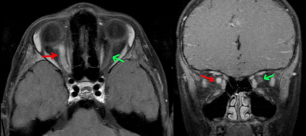
5 year old patient with 1 week of right eye blurry vision, then several days of right eye pain. Physical exam notable for right papilledema and progressively worsening vision....

This magazine is from a federated server and may be incomplete. Browse more on the original instance.
5 year old patient with 1 week of right eye blurry vision, then several days of right eye pain. Physical exam notable for right papilledema and progressively worsening vision....
This patient had episodic electric/shooting/radiating pain of the left face. An MRI was done....

[LEFT]: This patient had one of the longer cerebellar tonsillar herniations I’ve seen. The tonsil is peg-like in shape and extends quite far below the foramen magnum to the level of the C2 posterior arch. As a result, there is crowding at the foramen magnum that is enough to impede CSF flow, resulting in hydrocephalus with...

[LEFT]: The midbrain has a deep interpeduncular cistern, and the superior cerebellar peduncles are very prominent and elongated, making the brainstem at this level look like a molar tooth. This is a classic finding in Joubert syndrome....

Incidental finding of a superior lumbar hernia (Grynfeltt-Lesshaft hernia). In this case, only a lobule of retroperitoneal fat is herniating through the defect, but organs can also herniate through.

Continuing the theme of things extending into spaces they don’t belong in, this is an incidental finding of an inguinal hernia that contains a small portion of the bladder. The patient got the CT for other reasons....

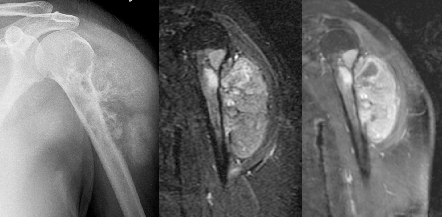
Female in her 30s with painful left shoulder....

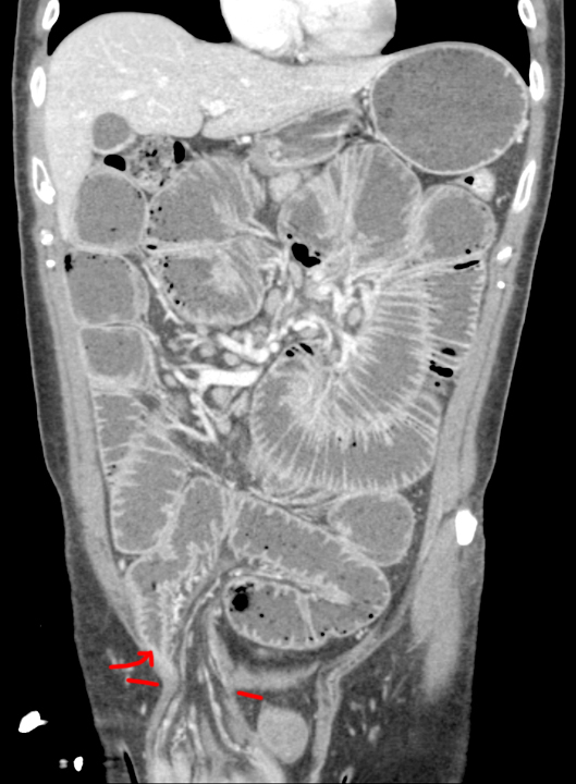
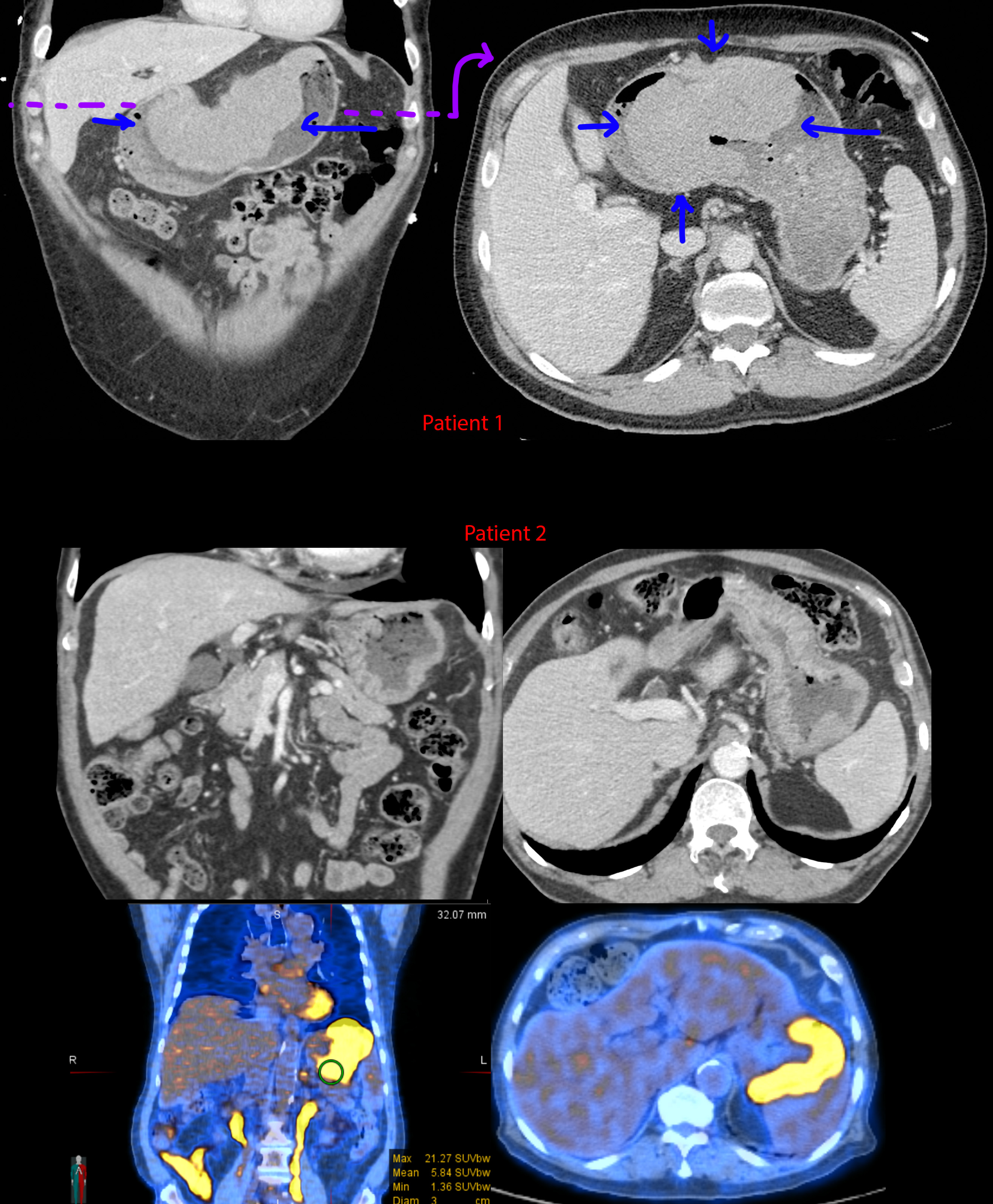
33 year old female with abdominal pain, abdominal distention, nausea/vomiting, early satiety, and weight loss....

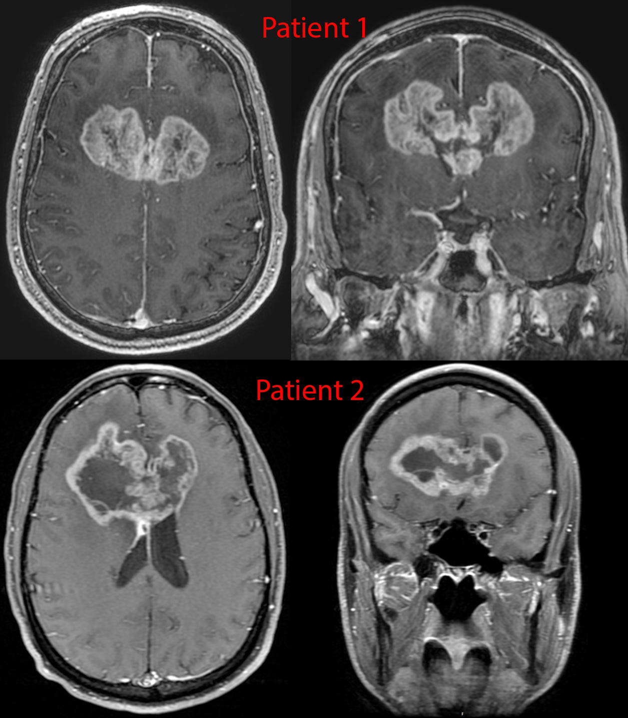
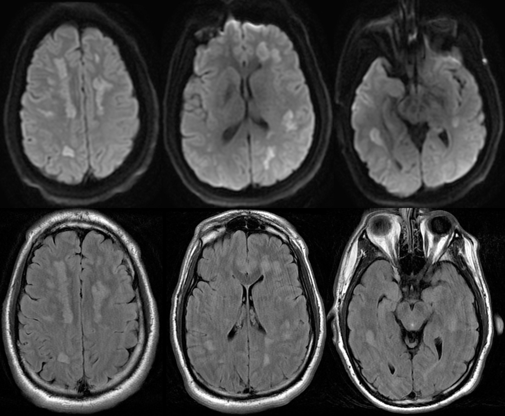
Postcontrast imaging of 2 patients with glioblastoma. These tumors are notorious for spreading along the white matter tracts - in this case the transverse fibers of the corpus callosum, given them a classic “butterfly” appearance.

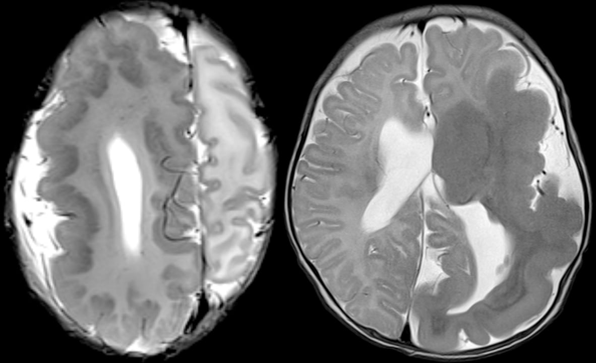
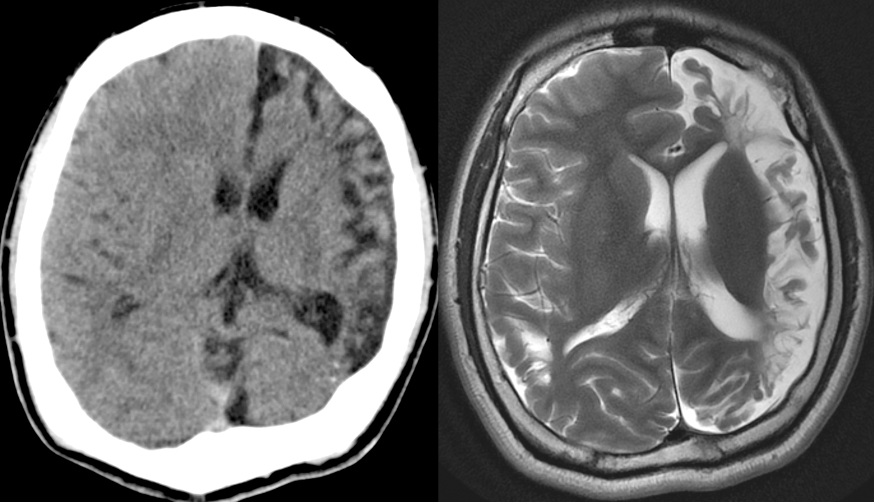
Two different patients with genetic disorders resulting in overgrowth of the brain....

Hello everyone!...
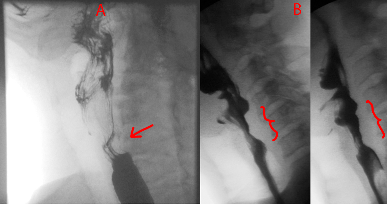
Quick one today. Take a look at Patient A and Patient B....

Patient was a young adult working in finance at a major tech company found to be mute and diaphoretic....

I remember this episode quite well because it happened around the time I decided to get into the medical field. In the episode, a young teacher had a first-time seizure while in the middle of teaching. House and team attempted to get a brain MRI, but she got an allergic reaction from the IV contrast. Thereafter, some drama...
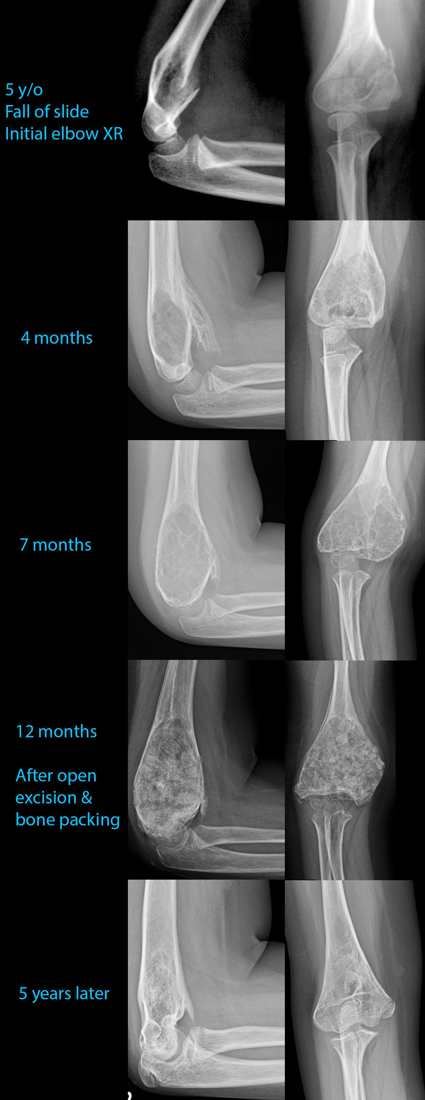
5 year old who fell off a slide....

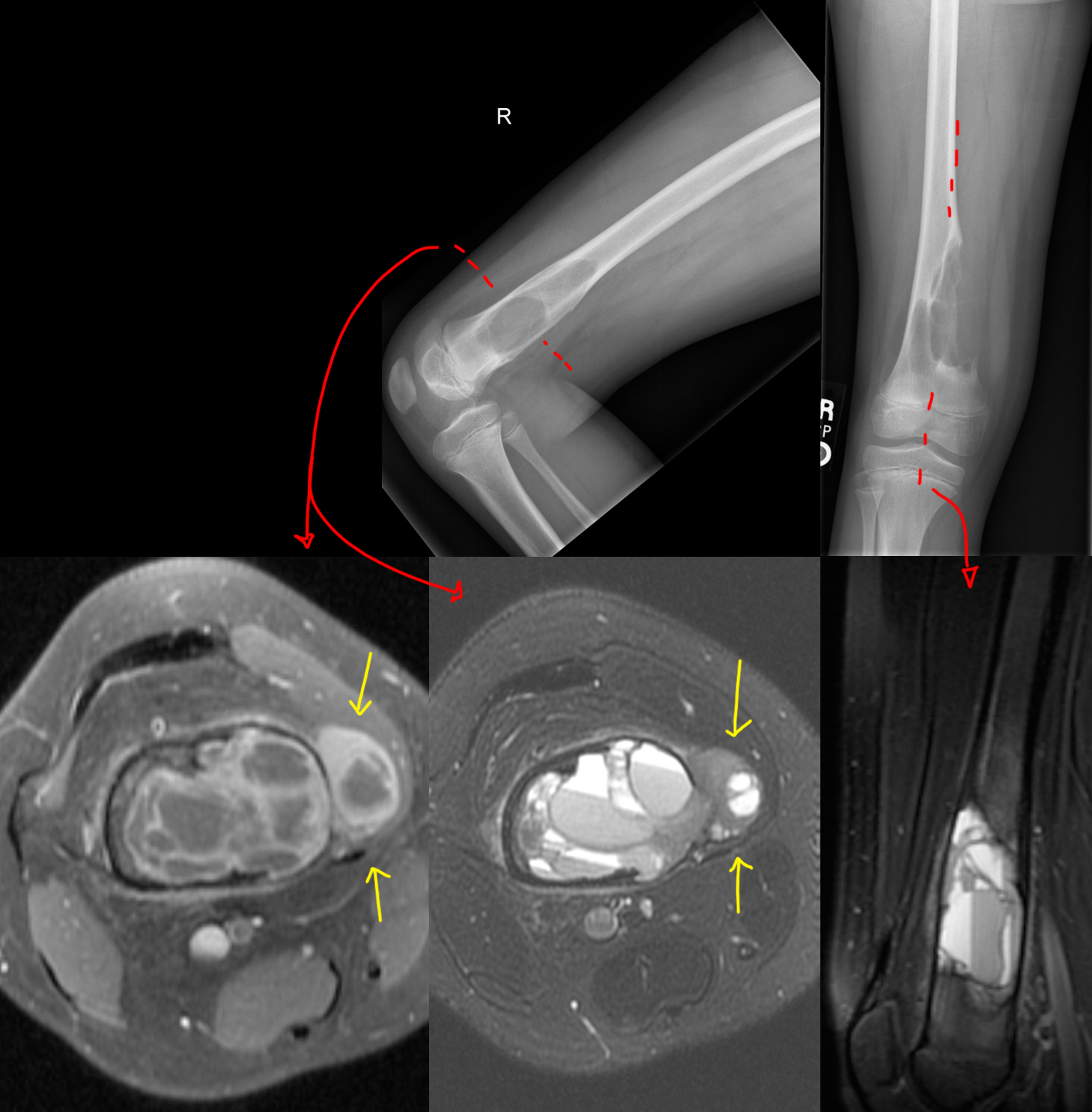
[Top]: X-ray shows a lucent, bubbly, lesion of the distal femur at the metaphysis. On the frontal view [top right], there is breakage through the medial femoral cortex into the adjacent soft tissues, not a good sign....

[Left]: Head CT shows left hemispheric volume loss. The injury happened early enough that even the skull is smaller on that side....

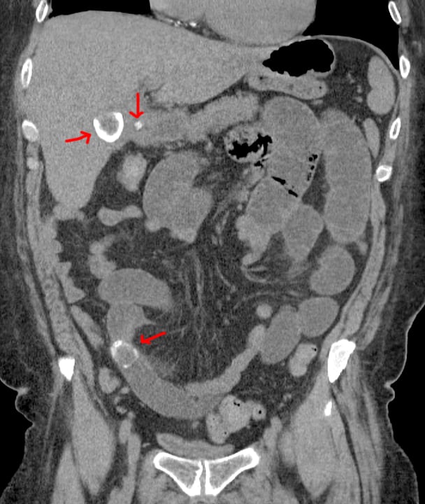
Red arrows point to 2 big gallstones, top one in the gallbladder and bottom one obstructing a small bowel loop, and a small gallstone in the cystic duct.

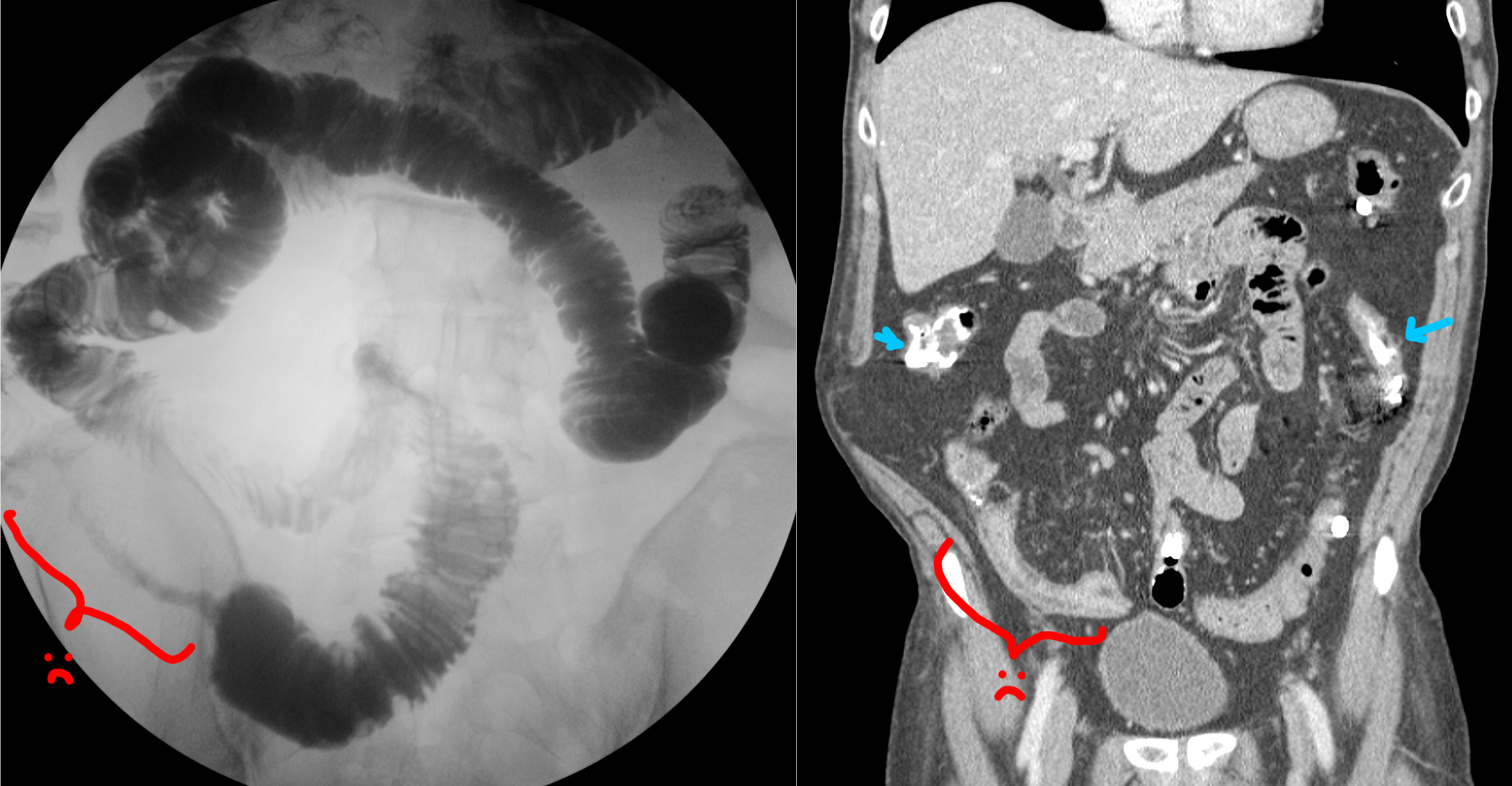
Red lines point to hernia entry. Red arrow points to where the bowel tapers and becomes obstructed as it enters the hernia sac.

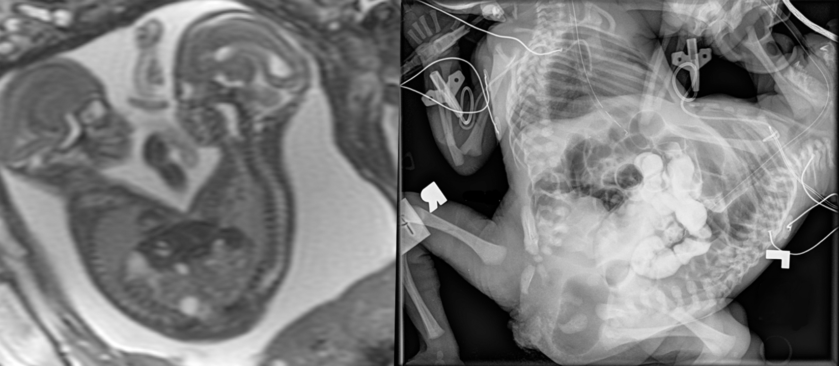
[Left]: Fetal MRI (FIESTA sequence) shows twins joined from their lower chest to the pelvis, but truly fused and sharing a single abnormal pelvic region. Not shown, but there are 3 lower limbs - one of the twins only had a single lower extremity....

No clinical history saved on this one - sorry....

I didn’t save any clinical history for these - sorry....


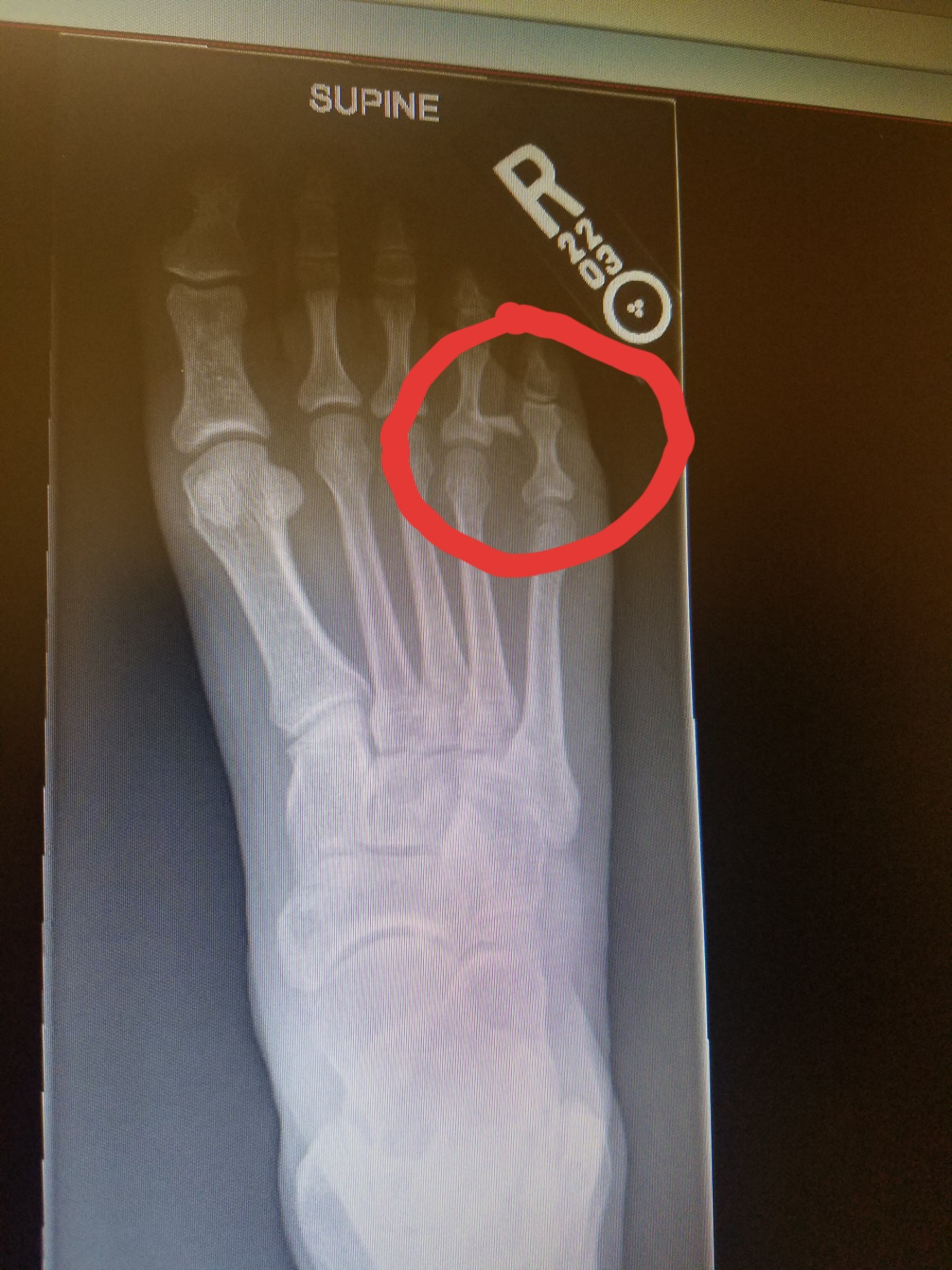
I can’t tell much from this xray, but that shit hurt for awhile

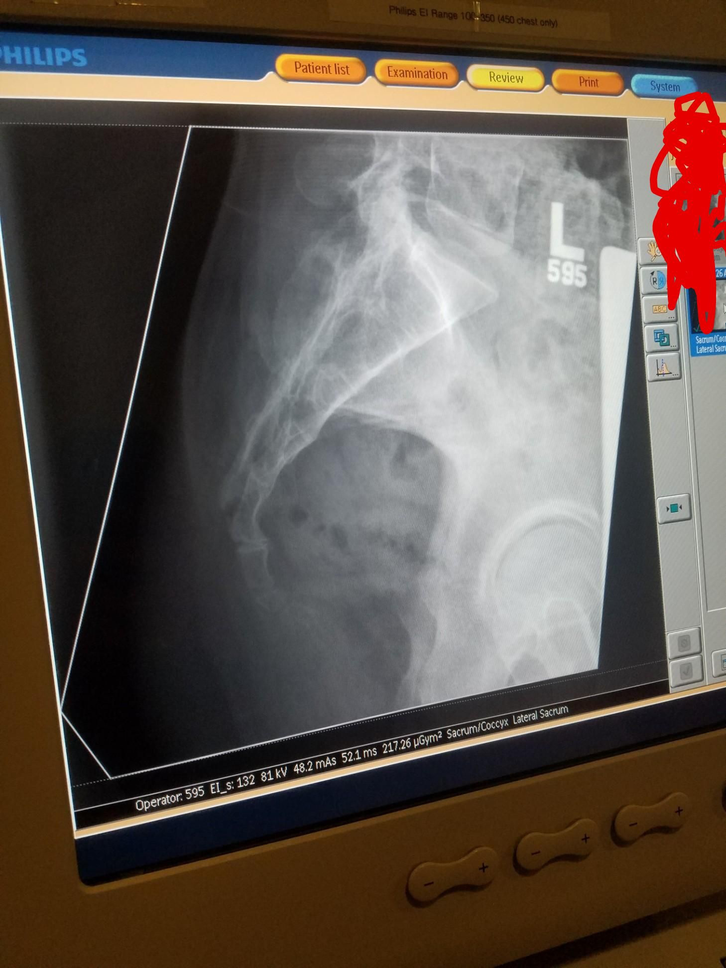
20-something year old male. Life will never be the same for him.
