jekely, New paper from Ikeda et al. on the biogenesis of chitin bristles in the annelid #Platynereis with nice #vEM reconstructions and a chitin synthase knockout.
Bristles are formed in a process of biological 3D printing. @biology
#microscopy
https://www.nature.com/articles/s41467-024-48044-3
steveroyle, Preprint describing Nellie, a napari plugin for automated organelle segmentation. Results look impressive!
https://arxiv.org/abs/2403.13214
repo (with video): https://github.com/aelefebv/nellie
Guinnessy, #AdaptiveOptics (AO) was developed to wipe out atmospheric aberrations from astronomical observations. The same technique is now being used in #microscopy to illuminate biological specimens.
teasingtabby, 
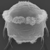
Microscopic dendrites of sodium hydrogencarbonate a.k.a. baking soda, viewed under a do-it-yourself smartphone-based microscope 🔬 in reflected white light.
Real width of the image is 2.7 mm 📏.
#microscopy #microscopyart #magnification #zoom #lens #chemistry #dendrite #dendrites #dendritic #hydrogencarbonate #bakingsoda #doityourself
FaithfullJohn, #ThinSectionThursday Plechroism in action: a wee gif showing pleochroic colour change in orthopyroxene (pale green-pink) + biotite (deep brown-pale brown) in this noritic rock from Govals, Aberdeenshire. Field of view about 2mm wide. #Microscopy #PolarizedLight #Pleochroism #Mineralogy #Geology
ScientistRachel, 🔬 🚨 Just 1 week left to apply to #IALSJanelia, @AICjanelia 's two-week long #microscopy bootcamp covering everything from #experimentalDesign to #imaging to #bioimageAnalysis
Read more and apply by Feb 15 here: https://www.aicjanelia.org/imagingacrosslengthscales
johannes_lehmann, Are you doing cell biology or biomedical research? Light microscopy-based imaging? If a facility for (multiplexed) imaging would pop up in your department, what would be your expectations and wishes for it? If you (would) run one, what is crucial?
I’ve applied for a (multiplexed) imaging facility manager job and would like to collect a few use scenarios (think tools, training, data analysis) to prepare for the interview.
#CellBiology #Microscopy #Imaging #Biology #LightMicroscopy #Science
AICjanelia, Reminder that there is 1 week left before the Feb 1 deadline to submit a proposal to use AIC #imaging technology and #microscopy expertise. Details at https://www.aicjanelia.org/apply
MWNautilus, German #Robotic #microscopy for everyone: The #OpenFlexure #OpenSource #microscope:
-#3Dprinted
-fully #automated using #RaspberryPi & #PiCamhttps://doi.org/10.1364/BOE.385729
#DIYbio #lab #instruments #imaging #OFM #fluorescence #MINT #STEM #education
furqanshah, 1/3; #Introduction 🕰️ I am an Associate Professor in Biomaterials @ University of Gothenburg, Sweden 🇸🇪 Research involves correlative #microscopy + #spectroscopy of mineralised tissues, e.g., bone 🦴🩻. Kickstart most mornings at the gym - because #fitness goals 💪🏻🏋️♂️! #academia #academicchatter #AcademicMastodon #Science #research #phd #mosstodon #icetodon #Fediverse #nature
ScientistRachel, My latest paper is now out in @J_Cell_Sci : https://journals.biologists.com/jcs/article/137/1/jcs261567/341218/Believing-is-seeing-the-deceptive-influence-of
Many thanks to my co-authors Leanna, Satya, Jesse, Leong, and to the entire @AICjanelia team for their feedback!
Every stage of a #microscopy project requires decisions, and those decisions are prone to #bias. In this paper, we use illustrative examples to describe potential pitfalls, and also provide strategies for mitigating bias.
You can also check out the data underlying our paper here: https://t.co/JqWOf5RedG
albertcardona, Course on "Quantitative Imaging: From Acquisition to Analysis"
Application deadline: January 31st
Course dates: April 1-16
Location: Cold Spring Harbor
Instructors: Jennifer Waters, @bethcimini @florianjug Hunter Elliott, Talley Lambert, Suliana Manley.
Financial aid is available to help offset tuition costs.
https://meetings.cshl.edu/courses.aspx?course=C-QICM&year=24
eLife, New #microscopy technology enables #nanoscale imaging of centimetre-scale tissues, making it possible to image the entire expanded mouse brain at cellular and subcellular resolution. #ReviewedPreprint #Neuroscience https://elifesciences.org/reviewed-preprints/91979?utm_source=mastodon&utm_medium=social&utm_campaign=organic
pixeltracker, #Janelia is offering a 12-day bootcamp designed to demonstrate how biological queries and hypotheses steer experimental designs on various #microscopy platforms and across length scales from molecules to small animals:
⏰ J uly 28 - August 9, 2024
⏰ Deadline to apply: Feb 15 (11:59 p.m. ET)
🌍 https://www.janelia.org/you-janelia/conferences/bootcamp-course-optical-imaging-across-biological-length-scales?Email_Preferences_Subscription_Center=&mc_cid=a9a3274574&mc_eid=fb2da94283#SuperResolutionMicroscopy #STED #LightSheetMicroscopy #ImageProcessing #DataAnalysis #FOSS #BigData
NikaShilobod, Hey @paleofire, I have a student that I am helping with his dissertation project and he needs to count #charcoal. Do you guys have any go to texts for lab and counting methods? I have not done this type of analysis before. #wildfire #palaeoecology #paleoecology #microscopy #sedimentology @geoscience
jekely, Nice review by Kirsty Wan and Rebecca Poon from Exeter on the diverse patterns and mechanisms of multiciliary coordination.
https://doi.org/10.1016/j.ceb.2023.102286
#cilia #microscopy #CellBiology #biophysics
albertcardona, The latest call for imaging proposals for the Advanced Imaging Center (AIC at #HHMIJanelia) is open.
Deadline is Feb. 1, 2024.
Get to use #HHMIJanelia imaging technology and expertise for free.
Visit http://aicjanelia.org/apply to schedule a technical consultation.
JulienDelpierre, French This is a render of a #microscopy image.
The rendering has been done on #blender using the tif to blender plugin developped by Oane Gros.
The tutorial is very well done and make it easy for beginners to get incredible results very quickly.Dev:
https://twitter.com/GrosOane
Plugin:
https://github.com/oanegros/tif2blender
Tutorial:
https://www.youtube.com/watch?v=TCQojYEYxVo
the_node, A while back we asked you, the Node community, to vote for your favourite artwork from @BSDB #DevBio_Art exhibition — the results are now in!
Have a look at the People's and Judges' choices of #microscopy images and #SciArt work:
https://thenode.biologists.com/peoples-choice-and-judges-choice-from-the-bsdb-the-node-virtual-art-exhibition/The exhibition is still open until the end of November! Have a stroll around:
🔬Scientific images
https://artspaces.kunstmatrix.com/node/12245599🖼#SciArt and 'Art by scientists'
https://artspaces.kunstmatrix.com/node/12245684🦞🐬🐢🐤Historic embryo zoo
https://artspaces.kunstmatrix.com/node/12186775
PPozzi, Oh, wow, what a warm reception! the fediverse science community may be small but is definitely welcoming!
As many suggested, here is my #introduction : I am a physicist developing optical microscopy methods for biomedical applications at the Politecnico di Milano University, in Italy. I will try to fill the fediverse with colorful images of squishy biological things. Apparently, I am also the new resident #opendata manager, so expect some posts on that!
#microscopy #lightsheet #optics
j_bertolotti, @PPozzi More optics on Mastodon is always a welcomed news! 😃
gsuberland, @PPozzi optics fedi is always awesome. lots of SEM/TEM nerds here too!
mariescopy, French 🇨🇵 12 months #microscopy assistant position in Bretagne, France:
CDD Assistant-e Ingénieur-e en #microscopie (12 mois) à la Station Biologique de #Roscoff
⚠️ Date limite de dépôt des candidatures 10 octobre ⚠️🔬 🦠 🌊
#marine #microalgae #macroalgae #protist #plankton #ecology
mzedp, This is not one photo, but 360 photos stitched together.
Each photo covering an area of around half a millimeter square.
This is just a small branch of some moss I picked up looking for Tardigrades.
moritz_negwer, #Jobalert: We are looking for a #protemics staff scientist for our team at the Ertürk lab at Helmholtz Munich! Want to work with a great team at the interface of microscopy and proteomics?
Check it out!
https://jobs.helmholtz-muenchen.de/jobposting/84eac3115b49ff2ecfc8f708f4fd598b3a9cf9320?ref=homepage
philpem, (edited ) Comparison of Brightfield versus DIC illumination, Motorola 68HC05 microprocessor.
The quality here is fairly poor; my light source polariser is a piece of cheap polarising film clipped into a 3D-printed frame.
I need to find somewhere that sells good quality ~18 to ~22mm glass polarizers.
#ICRE #microscopy #reverseengineering